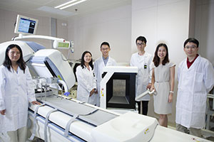
A picture is worth a thousand words. This is clearly true in medical diagnosis. Indeed, medical imaging has become a powerful tool in the diagnosis and treatment of illness. Over the past seven years, the team in the Biomedical Imaging Laboratory, established by the Faculty of Science and Technology (FST), has worked relentlessly in this field, producing encouraging results that have garnered international recognition. Prof Greta Mok, founder of the laboratory, hopes to apply artificial intelligence (AI) and other related technologies in nuclear medicine to improve diagnosis and treatment of medical conditions for Macao residents.
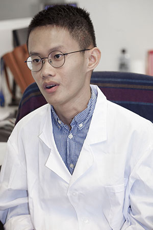
Prof Mok is an associate professor in the Department of Electrical and Computer Engineering and a joint associate professor in the Faculty of Health Sciences. She is the first native of Macao to become a medical imaging scholar in Macao. At age 18, she left Macao to pursue higher education at Yang-Ming University in Taiwan. After graduation, she was admitted by Johns Hopkins University in the United States, a universally acclaimed institution in the fields of medicine and public health. She is also the first Macao native to obtain a doctoral degree at JHU. She was later recruited by the Chinese University of Hong Kong as a junior faculty member before returning to Macao to join UM in 2010. Two years later, in 2012, she founded Macao’s first Biomedical Imaging Laboratory.
Nuclear Medicine Imaging
In nuclear medicine, patients are given small amounts of radioisotope-labelled drugs either orally or by injection. The radioisotopes then circulate through the body and are absorbed specifically by target organs or tumours under examination. The isotopes undergo radioactive decays and emit gamma rays from within the body, which are captured by a gamma camera to create images. By reconstructing and analysing these images, doctors can assess organ functions, tumour location, disease progression and staging, and effectiveness of the treatment. Prof Mok says that organ movements, such as the beating of the heart or the inflation and exhalation of the lungs, can sometimes lead to artifacts, which can degrade the interpretations of imaging results.
The research team currently focuses on the development of nuclear medical instrumentation, methods of medical image generation, and subsequent processing and analysis, with the aim of making medical images of the brain, heart, liver and other organs more accurate, in order to improve the medical diagnosis and treatment of cancers. ‘Take the brain for example. We can’t directly assess the brain functions from the physical examination,’ says Prof Mok. ‘There are a lot of diseases such as Parkinson’s disease and dementia that don’t have obvious symptoms before the onset of the diseases. So the best way to detect disease pre-emptively is to use non-invasive medical imaging techniques, and nuclear medicine has obvious advantages over other imaging technologies in this application.’ In the assessment of a tumour, the doctor can prescribe a positron emission tomography (PET) scan, where glucose labelled with radioactive fluorine is injected into the patient’s body. Since the malignant tumour usually absorbs more glucose than normal tissues, the doctor can pinpoint the location of the tumour and assess the severity of the condition by detecting and quantifying the unusually ‘bright spots’.
Three Major Research Projects
According to Prof Mok, the lab currently has three major research projects. The first is the development of an active breathing controller to remove respiratory artefacts and related image acquisition and post-processing software. When the patient is scanned, this controller can suspend the patient’s breathing while collecting images, thereby removing respiratory artefacts, improving the quality of imaging, and enabling more accurate quantification of the lesions. The second project is to design and develop a multi-purpose, multi-pinhole collimator that can collect more gamma photons than traditional single-pinhole and parallel-hole collimators. This collimator can enhance the sensitivity of the scanner, which can in turn reduce image noise, radiation dose or scanning time. The lab has commissioned a Dutch company to customise a prototype of the collimator. The device has already been installed on the lab’s single-photon computed tomography (CT) scanner and is undergoing further evaluation.
In addition to diagnosis, radioactive drugs can also be used for treatment of cancers, which is exactly the focus of the lab’s third project. The lab has developed a piece of 3D computer software for internal dose calculation in targeted radionuclide therapy. It can be used to evaluate the radiation dose absorbed by the tumour and various organs, an important index for treatment efficacy and potential toxicity. Hospitals and research institutions can apply this software to optimise dose calculation for each individual patient, so that treatment planning could be more precise and personalised.
Prof Mok says that AI technology is increasingly used in medical imaging research. She says, ‘AI technology not only can help to improve the image quality, it can also reduce the radiation dose. I hope to apply AI technology in all three research projects. With our first project on respiratory artifact reduction, we have already successfully reduced image noise using AI technology. In the future, we will study the use of AI technology to reduce the radiation dose and image acquisition time to benefit the patients.’
Enormous Potential
Under the leadership of Prof Mok, the lab has trained over a dozen postgraduate students from Macao and mainland China, who are now working at universities, industries, and medical institutions in Macao and other parts of the world. Among them, doctoral students Zhang Duo and Sun Jingzhang foresee enormous potential in applying AI technology to medical imaging research.
Over the past few years, under the guidance of Prof Mok, Zhang has published some papers on medical image correction, including one paper that examines the effects of different respiratory patterns on medical images. During his doctoral study, Zhang was recommended by Prof Mok to work as a visiting scholar for one year at the University of Massachusetts Medical School and Yale University in the US, where he participated in cutting-edge research. ‘I gained a lot from working at Yale. According to my supervisor at Yale, I did in one month what other students did in one semester,’ Zhang says. Because of his outstanding performance and the solid grounding in research he developed at UM, Yale’s supervisor invited him to be a postdoctoral research fellow at the university, but he had other plans. ‘Our lab is in close contact with research institutions in the Greater China region, Europe, and the US, and we also collaborate with institutions in the Greater Bay Area, such as the Chinese University of Hong Kong and Shenzhen Institutes of Advanced Technology of the Chinese Academy of Sciences. I plan to find a job at a university in the Greater Bay Area after graduation, because I see great potential for career development in this area. It would also be more convenient for me to keep in close research contact with Prof Mok.’
Sun Jingzhang, another of Prof Mok’s PhD students, has both a bachelor’s and master’s degree in computer science. One of his research projects at UM involves using artificial neural networks to reduce noise in medical imaging. ‘I have a background in computer science, and I used to study image processing. There aren’t many people doing research in this area, so I want to explore more,’ he says. Sun now uses deep learning techniques to try to tackle some challenging issues concerning medical image quality.
International Recognition
The research results from the lab have been applied on more than 50 patients in Taiwan and the US to improve their medical diagnosis. The team has also won many awards at international conferences in recent years. At the Third Asian Nuclear Medicine Academic Forum in 2017, the team stood out among strong research groups from Asia and won the sole first prize in the Fourth Rising Nuclear Medicine Professional Challenge, the highest honour of the forum, for their work titled ‘A Framework for Improved 3D Personalised Targeted Radionuclide Therapy Dosimetry Using Registration on Sequential ECT/CT’. It was the first time that a team from Macao had won this award. The same project later received the International Best Abstract Award and the third prize of the Computer and Instrumentation Council (CalC) Young Investigator Award (YIA) at the 64th Annual Meeting of the Society of Nuclear Medicine and Molecular Imaging held in Denver, Colorado, US in the same year. It was the first time a research team from the Greater China region had been shortlisted for, much less received, an award in this YIA category. Two other notable teams shortlisted for this award were from Stanford University and MD Anderson Cancer Center.
The Year 2018 continued to be a fruitful year for Prof Mok’s team. At the 65th Annual Meeting of the Society of Nuclear Medicine and Molecular Imaging (SNMMI) held in Philadelphia, Prof Mok received the International Best Abstract Award for the second consecutive year for her work titled ‘Comparison of Different Tc-99m-MAA Imaging Protocols for Y-90 SIRT Treatment Planning’, as well as the Tracy Lynn Faber Memorial Award, in recognition of her major contributions to high performance Emission Computed Tomography and Computed Tomography instrumentation, reconstruction, and analysis. She is the first Chinese scientist to ever receive this prestigious award.
‘These international awards are certainly very encouraging for the research team,’ says Prof Mok. ‘In a “desert of isotopes” like Macao, it is exceedingly difficult to conduct research in nuclear medicine. For the same experiment, you need to work extra hard to make it happen. Most clinical trials have to be conducted outside of Macao. All the past winners of these international awards had the full support of their medical schools. UM doesn’t have a medical school, and isotopes are just hard to obtain. But the team managed to overcome all the difficulties and won these international awards. We feel really honoured and thankful.’
It is not easy to build a lab from scratch. Prof Mok says, ‘I am very grateful to my team and my students for their support. I am also grateful to the university for its trust and to the international and local societies for their recognition. Because of them, our hard work over the years has not been in vain. Everyone in the lab puts their hearts and souls into what they are doing. They are full of dreams and they are passionate about their research. I hope the fruits of our research can be applied in Macao as well. Hopefully we will increase collaborations with hospitals in Macao to let local patients be the first to benefit from our technology. Our goal is to bring real changes in the diagnosis and treatment of illnesses for local residents using medical imaging technologies.’
網絡有云,有圖有真相,醫學影像也成為疾病診治不可或缺的一環。為了這個目標,澳門大學科技學院的生物醫學影像實驗室經過七年來不斷努力,近年初見成果,屢獲國際獎項。實驗室始創人莫昇萍教授說,希望深化運用人工智能等技術,進一步改良核醫學診斷和治療,改善巿民健康。
莫昇萍是科技學院電機及電腦工程系副教授、健康科學學院兼任副教授,澳門首位土生土長的醫學影像學者。莫教授18歲離開澳門到台灣陽明大學升學,畢業後負笈到在醫學與公共衛生領域享譽全球的美國約翰•霍普金斯大學攻讀博士,是該學院首位來自澳門的博士畢業生。她之後曾在香港中文大學任教,2010年回流澳門加入澳大,2012年創立澳門首個生物醫學影像實驗室。
核子醫學影像
在核醫學診斷方面,病人會被注射或口服少量放射性藥物(同位素)。這些同位素會分佈在人體不同的部位,具特異性的積累到待診斷的器官或腫瘤,並放出伽馬射線到人體以外,被專門的伽馬照相機收集和生成影像。通過重建並分析這些影像,專家就能觀察器官的功能、腫瘤的位置、嚴重程度和有否轉移,並且評估治療效果等。莫教授說,器官移動(例如心跳或肺部充氣呼氣)有時會令醫學影像出現假影,影響診斷。
研究團隊主要研究核醫學儀器的開發、醫學影像生成的方法和後續的處理分析,讓醫學影像更加精準,涉及大腦、心臟、肝臟等多個器官,以及癌症的診斷和治療。莫教授說:「譬如說腦部,我們無法直接觀察腦部的功能,而柏金遜症、失智症等不少病症發病前都沒有明顯症狀,最好的檢查方法,就是非侵入性的醫學影像掃瞄,而在此應用上,核醫學又比其他影像技術具有明顯優勢。」此外,偵測與定性腫瘤時,醫護人員通常會安排患者接受正電子掃描,先將有放射性氟標記的葡萄糖注射到體內。由於腫瘤會吸收很多葡萄糖,只要觀察不正常的「亮點」,就能得知腫瘤的位置和嚴重程度,以及有沒有潛在的轉移。
三大研究項目
莫教授說,實驗室目前有三個主要的研究項目。「第一個是去除呼吸假影的呼吸控制器、相關影像採集及後期處理軟體的開發。病人接受掃描時,這個呼吸器可以主動地控制病人呼吸,然後採集影像,從而去除呼吸假影,提升對病灶的偵測能力和量化精度。」第二個項目則是設計和製造一台多用途的多針孔準值儀。該儀器比傳統的單針孔與平板準值儀採集到更多伽馬光子,增強掃描儀的靈敏度,繼而用以降低影像噪聲、減低輻射劑量或縮短掃描時間。實驗室已經委託荷蘭一家公司訂製該準值儀的原形,並安裝在實驗室的單光子電腦斷層掃描儀上再作評估。
除了診斷,放射性藥物也能用於治療,實驗室的第三個項目就能為接受標靶核素治療的病人計算藥物劑量,以及評估藥物對腫瘤的療效和各個器官帶來的放射劑量。「我們研發出一個三維核醫同位素治療劑量計算軟體,可供醫院或研究機構採用,優化劑量計算,令每個病人都有更準確、更個人化的注射劑量計算和輻射劑量評估。」
莫教授說,運用人工智能是醫學影像研究的一個趨勢。「人工智能可以提高影像質素,也能減少輻射劑量,我希望三個研究項目都能應用上人工智能技術。在去除呼吸假影的項目上,我們已經成功應用人工智能減低影像的噪聲。我們將來也會研究用人工智能,減少掃描所需的同位素劑量或生成影像的時間。」
發展潛力
在莫教授帶領下,實驗室培養了十多位來自澳門和內地的研究生,目前在世界各地及本澳的院校、業界和醫療機構發光發熱,其中在讀博士生張鐸和孫敬張看見醫學影像研究與人工智能結合的前發展前景。
過去數年,張鐸在莫教授指導下,發表了不少關於醫學影像校正的論文,包括涉及不同呼吸模式對醫學影像的影響。攻讀博士期間,張鐸獲莫教授推薦,到美國麻省大學醫學院和耶魯大學擔任訪問學者一年,參與前沿研究。「在耶魯工作的收穫很大,根據那邊導師的評價,我一個月做了別人一個學期才做完的工作。」由於表現出色和在澳大培養出的研究根基,耶魯大學的導師邀請張鐸到該校做博士後研究,但他認為畢業後留在粵港澳大灣區發展也是一個理想的選擇。「我們實驗室與大中華地區和歐美的科研機構聯繫密切,跟香港中文大學和中國科學院深圳先進技術研究院等大灣區院校也有合作,畢業後到大灣區其他大學任職發展潛力大,還方便以後跟莫教授在研究上保持緊密聯繫。」
莫教授另一位博士生孫敬張的本科和碩士都是計算機科學。他在澳大的其中一項研究是運用人工神經網絡技術,降低醫學影像的噪聲。「首先我有計算機科學背景,之前也是做圖像處理。由於從事這方面研究的人也較少,所以我想嘗試探索更多。」孫敬張現在用深度學習技術,解決一些關於醫學影像質素的難題。
國際肯定
實驗室的成果已在台灣和美國超過50個病人身上得到應用,提高了醫學影像的質素。團隊近年更在國際會議奪得多個獎項。在2017年的第三屆亞洲核醫學論壇上,團隊以研究課題「兼備圖像對位算法的三維個人化靶向核素治療劑量計算架」,力壓一眾亞洲隊伍,榮獲論壇最高榮譽獎項「第四屆亞洲青年競賽」唯一一等獎,是首支來自澳門的獲獎隊伍。相關項目同年再下一城,在美國丹佛巿舉行的北美核醫學與分子影像年會獲頒「國際最佳論文獎」和「青年研究家獎」第三名。其中在競逐「青年研究家獎」方面,實驗室是儀器與軟件類別首支來自大中華地區的入選團隊,力壓史丹福大學和德州安達森癌症中心的世界頂級強隊獲獎,讓澳門核醫研究在國際舞台上一鳴驚人。
在2018年北美核醫學與分子影像年會上,莫教授憑著針對治療肝臟腫瘤的研究再奪「國際最佳論文獎」。大會更向她頒發專門表揚傑出女科學家的「Tracy Lynn Faber MemorialAward」,表彰其對射線斷層成像與計算機斷層成像的設備、圖像重建和分析研究等的出色貢獻,令莫教授成為首名得獎的華人科學家。
莫教授說,這些國際肯定都是對研究團隊的莫大鼓舞。「要在澳門這個『同位素沙漠』從事核醫學研究異常艱鉅,同一項實驗要比其他地方付出多幾倍努力才能實現,臨床測試則大多要到外地進行。一眾國際獎項過去的得獎者都有醫學院的全力支持,澳大沒有醫院,而且難以取得同位素,但團隊也能排除萬難,得到國際認可,實在非常榮幸。」
Prof Mok’s research team focuses on the development of nuclear medical instrumentation, methods of medical imaging generation, and subsequent processing and analysis techniques.
莫昇萍教授的研究團隊主要研究核醫學儀器的開發、醫學影像的生成與後處理
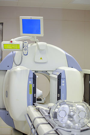
The lab has a SPECT/CT scanner, which was donated by Kiang Wu Hospital
實驗室有一台由澳門鏡湖醫院所贈的複合式單光子發射電腦斷層掃描
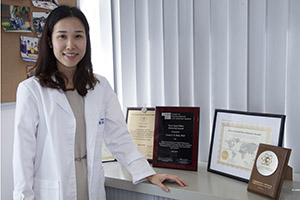
Prof Greta Mok’s team has won many international awards
莫昇萍教授和團隊的研究屢獲國際殊榮
During his doctoral study, Zhang Duo was recommended by Prof Mok to work as a visiting scholar for one year at the University of Massachusetts Medical School and Yale U, USA
張鐸讀博期間獲薦到美國麻省大學醫學院和耶魯大學擔任訪問學者一年
Sun Jingzhang is working on the application of artificial neural networks to reduce noise in medical imaging under the guidance of Prof Mok.
孫敬張在莫昇萍教授指導下,利用人工神經網絡技術,降低醫學影像的噪聲。